|
|
|
Research
Study of the mechanisms in low-frequency oscillation
of the resting state brain
|
Low-frequency oscillation of the resting state brain |
|
|
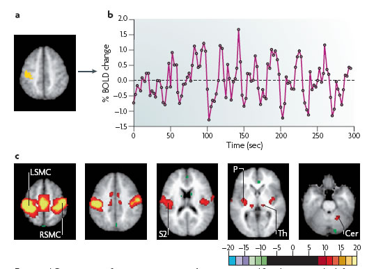
Recently, there has been a growing interest about the default mode network and resting state network analysis by correlating low-frequency fluctuations in BOLD signals, and the changes in these networks with neurological disease.
The spontaneous BOLD fluctuations were not random noise and specifically correlated between functionally related brain regions. The fluctuations of BOLD signal also well correlate with fluctuations of high frequency neural activity. And the spontaneous fluctuations do not disappear during task conditions and this may be due to contribute to inter-trial variability. |
Research Purpose
• To elucidate the biological mechanism and the role of low-frequency oscillations of resting state fMRI BOLD signal
• To prove if the BOLD signal low-frequency oscillations are due to the secondary effect caused by neuronal activity of intrinsic periodic vessel contraction
Expected Contribution
For understanding of the dynamics of brain operation and improvement of
• Medical science and pharmacology
• The field of brain engineering
(1) Intrinsic Optical Signal (IOS) Imaging
Detection of cerebral blood volume or relative concentration of total and reduced hemoglobin in living mice
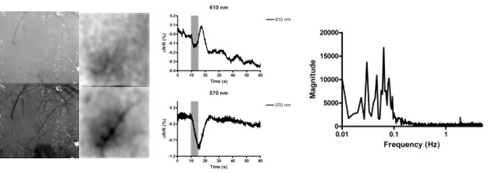 (Left) Reference image and response map of IOSI on the somatosensory cortex of living mouse, (Center) Relative reflectance changes of total and reduced hemoglobin by electrical hindpaw stimulation showing CBV changes, (Right) Low-frequency oscillation observed at below 0.1 Hz
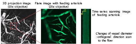
(2) Two-Photon Laser Scanning Microscopy
In Vivo brain imaging experiment for observation of structural and functional cerebral blood vessel changes
(3) Local Field Potential
Measurement of neural activity linked with hemodynamic responses with large spatial resolution
|
|
|
|